Pneumothorax - Causes, Symptoms, Diagnosis, Treatment & Prevention
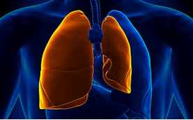
Pneumothorax is a condition of lung collapse. Pneumothorax occurs when air enters her space in the lung area (pleural space). The air can enter the pleural space when there are open sores on the chest wall - torn or ruptured lung tissue (pulmonary bullae), this disturbs the pressure that makes the lungs bulge.
Treatment of pneumothorax is usually done by inserting a needle or chest tube between the ribs to remove excess air. However, a small pneumothorax can heal by itself.
Risk factors for pneumothorax are:
Examples of injuries that can cause traumatic pneumothorax are:
Chest trauma due to motor vehicle accidents
Rapid treatment of pneumothorax due to significant chest trauma is very important. The symptoms are often severe, and they can contribute to potentially fatal complications such as cardiac arrest, respiratory failure, shock, and death.
There are two main types of spontaneous pneumothorax: primary and secondary. Primary spontaneous pneumothorax (PSP) occurs in people who do not have lung disease, often attacking tall and thin young men. Secondary spontaneous pneumothorax (CNS) tends to occur in older people with known lung problems.
Some conditions that increase the risk of secondary spontaneous pneumothorax include:
Other symptoms of pneumothorax may include:
The imaging tests commonly used to diagnose pneumothorax are:
Treatment options can include close observation combined with chest tube insertion, or a more invasive surgical procedure to resolve and prevent further lung collapse. Oxygen can be given.
Regular physical activity has not been shown to worsen or delay healing of pneumothorax. However, it is often recommended that intense physical activity or high contact sports be delayed until the lungs heal completely and the pneumothorax disappears.
Pneumothorax can cause a decrease in oxygen levels in some people. This condition is called hypoxemia. If this is the case, the doctor will order oxygen supplementation along with restrictions on activity.
Needle aspiration may be less comfortable than chest tube placement, but it is also more likely to need to be repeated.
For the installation of a chest tube, the doctor will insert a hollow tube between the patient's ribs. This allows air to flow and the lungs to rise again. The chest tube may remain in place for several days if there is a large pneumothorax.
During pleurodesis, the doctor irritates the pleural space so that air and fluid no longer accumulate. The term "pleura" refers to the membrane that surrounds each lung. Pleurodesis is done to make the lung membrane stick to the chest cavity. After the pleura attaches to the chest wall, the pleural space no longer expands, and this prevents the formation of pneumothorax in the future.
Mechanical pleurodesis is done manually. During surgery, the surgeon brushes the pleura to cause inflammation. Chemical pleurodesis is another form of treatment. The doctor will send irritant chemicals to the pleura through the chest tube. Irritation and inflammation causes the pleural lungs and layers of the chest wall to stick together.
There are several types of surgery for pneumothorax. One option is thoracotomy. During this operation, the surgeon will make an incision in the pleural space to help them see the problem. After the surgeon performs a thoracotomy, they will decide what to do to help the patient recover.
Another option is thoracoscopy, also known as video Thoracoscopic Surgery (VATS). The surgeon inserts a small camera through the patient's chest wall to help them look into the chest. Thoracoscopy can help surgeons decide on treatment for pneumothorax. Possibilities include sewing closed blisters, closing air leaks, or removing collapsed parts of the lungs, called lobectomy.
Some tips for preventing pneumothorax recurrence are:
Also Read :
What is a pneumothorax?
Pneumothorax is a lung condition caused by blunt or penetrating chest injuries, certain medical procedures, or damage from lung disease. This disease may occur for no apparent reason. Symptoms of pneumothorax usually experience sudden chest pain and shortness of breath. In some cases, collapsed lungs are at risk of death.Treatment of pneumothorax is usually done by inserting a needle or chest tube between the ribs to remove excess air. However, a small pneumothorax can heal by itself.
Causes of pneumothorax
The following are some of the conditions and diseases that cause pneumothorax are:1. Chest injury
Blunt or penetrating injuries to the chest can cause lung collapse. Some injuries or pulmonary bulls can occur due to injuries due to car collisions, while other cases may accidentally occur when a medical procedure is used that uses a needle into the chest.2. Lung disease
Damaged lung tissue is more likely to collapse. Lung damage can be caused by several types of diseases, including chronic obstructive pulmonary disease (COPD), cystic fibrosis, and pneumonia.3. Blisters
Small air blisters (blebs) can develop in the upper part of the lungs. This bleb sometimes breaks - allowing air to leak into the space surrounding the lungs.4. Mechanical ventilation
A severe type of pneumothorax can occur in people who need mechanical handling to breathe. A ventilator can be used to create an imbalance of air pressure inside the chest. The thing to be aware of is that the lungs can collapse completely.Risk Factors for Pneumothorax
Men are far more likely to experience pneumothorax than women. The type of pneumothorax caused by blisters caused by ruptured air is most likely to occur in those between the ages of 20 and 40 years, especially if the person has a tall and thin posture.Risk factors for pneumothorax are:
1. Smoking
The risk can increase by how long and how many cigarettes are smoked, even without emphysema - the air sacs in the lungs can be damaged.2. Genetics
Certain types of pneumothorax may appear due to offspring from a family history.3. Lung disease
Experiencing lung disease - especially chronic obstructive pulmonary disease (COPD) - which might make the lungs collapse.4. Mechanical ventilation
People who need mechanical ventilation to help their breathing may be at a higher risk of developing a pneumothorax.5. Previous pneumothorax
Someone who has had one pneumothorax will be at higher risk of experiencing another type of pneumothorax.Type of pneumothorax
The entry of air into the pleural cavity can be divided into two ways, namely traumatic pneumothorax and nontraumatic pneumothorax:1. Traumatic pneumothorax
Traumatic pneumothorax occurs after several types of trauma or injury occur in the chest wall or lungs. This condition can be a small or significant injury. Trauma can damage the structure of the chest and cause air to leak into the pleural space.Examples of injuries that can cause traumatic pneumothorax are:
Chest trauma due to motor vehicle accidents
- Broken ribs
- A hard blow to the chest from contact sports, such as from soccer tackles
- Stab wounds or gunshot wounds to the chest
- Medical procedures that can damage the lungs, such as central channel placement, ventilator use, lung biopsy, or CPR
Rapid treatment of pneumothorax due to significant chest trauma is very important. The symptoms are often severe, and they can contribute to potentially fatal complications such as cardiac arrest, respiratory failure, shock, and death.
2. Nontraumatic pneumothorax
This type of pneumothorax does not occur after injury. Namu, this condition occurs spontaneously, which is why it is also called spontaneous pneumothorax.There are two main types of spontaneous pneumothorax: primary and secondary. Primary spontaneous pneumothorax (PSP) occurs in people who do not have lung disease, often attacking tall and thin young men. Secondary spontaneous pneumothorax (CNS) tends to occur in older people with known lung problems.
Some conditions that increase the risk of secondary spontaneous pneumothorax include:
- Chronic obstructive pulmonary disease (COPD), such as emphysema or chronic bronchitis
- Acute or chronic infections, such as tuberculosis or pneumonia
- Lung cancer
- Cystic fibrosis , a genetic lung disease that causes mucus to accumulate in the lungs
- Asthma, a chronic obstructive airway disease that causes inflammation
Symptoms of pneumothorax
Symptoms of traumatic pneumothorax often appear during trauma or chest injury, or not long after that. While the onset of symptoms of spontaneous pneumothorax usually occurs at rest. The sudden onset of chest pain is often the first symptom.Other symptoms of pneumothorax may include:
- Stable pain in the chest
- Shortness of breath, or dyspnea
- Cold sweat
- Tightness in the chest
- Blue, or cyanosis
- Severe tachycardia, or rapid heartbeat
Diagnosis of pneumothorax
The diagnosis is based on the presence of air in the space around the lungs. The stethoscope can detect changes in lung sound, but detecting a small pneumothorax may be difficult. Some imaging tests may be difficult to interpret because of the position of the air between the chest wall and the lungs.The imaging tests commonly used to diagnose pneumothorax are:
- Upright posteroanterior chest radiograph
- CT scan
- Thoracic ultrasound
Pneumothorax treatment
Treatment and treatment will depend on the severity of the patient's condition. This will also depend on whether the patient has had a previous pneumothorax and what symptoms have been experienced. For treatment, surgical and non-surgical treatments are available.Treatment options can include close observation combined with chest tube insertion, or a more invasive surgical procedure to resolve and prevent further lung collapse. Oxygen can be given.
1. Observation
Observation or waiting is usually recommended for those who have a small PSP and who are not out of breath. In this case, the doctor will monitor the patient's condition regularly because air absorbs from the pleural space. Frequent X-rays will be taken to check if the patient's lungs have fully developed again. The doctor will likely instruct the patient to avoid air travel until the pneumothorax is completely finished.Regular physical activity has not been shown to worsen or delay healing of pneumothorax. However, it is often recommended that intense physical activity or high contact sports be delayed until the lungs heal completely and the pneumothorax disappears.
Pneumothorax can cause a decrease in oxygen levels in some people. This condition is called hypoxemia. If this is the case, the doctor will order oxygen supplementation along with restrictions on activity.
2. Reducing excess air
Needle aspiration and chest tube insertion are two procedures designed to remove excess air from the pleural space in the chest. This can be done at the bedside without the need for general anesthesia.Needle aspiration may be less comfortable than chest tube placement, but it is also more likely to need to be repeated.
For the installation of a chest tube, the doctor will insert a hollow tube between the patient's ribs. This allows air to flow and the lungs to rise again. The chest tube may remain in place for several days if there is a large pneumothorax.
3. Pleurodesis
Pleurodesis is a more invasive form of pneumothorax. This procedure is generally recommended for individuals who have experienced episodes of recurrent pneumothorax.During pleurodesis, the doctor irritates the pleural space so that air and fluid no longer accumulate. The term "pleura" refers to the membrane that surrounds each lung. Pleurodesis is done to make the lung membrane stick to the chest cavity. After the pleura attaches to the chest wall, the pleural space no longer expands, and this prevents the formation of pneumothorax in the future.
Mechanical pleurodesis is done manually. During surgery, the surgeon brushes the pleura to cause inflammation. Chemical pleurodesis is another form of treatment. The doctor will send irritant chemicals to the pleura through the chest tube. Irritation and inflammation causes the pleural lungs and layers of the chest wall to stick together.
4. Operations
Surgical treatment for pneumothorax is needed in certain situations. Patients may need surgery if they have had a recurrent spontaneous pneumothorax. The amount of air trapped in the chest cavity or other lung conditions can also guarantee surgical repair.There are several types of surgery for pneumothorax. One option is thoracotomy. During this operation, the surgeon will make an incision in the pleural space to help them see the problem. After the surgeon performs a thoracotomy, they will decide what to do to help the patient recover.
Another option is thoracoscopy, also known as video Thoracoscopic Surgery (VATS). The surgeon inserts a small camera through the patient's chest wall to help them look into the chest. Thoracoscopy can help surgeons decide on treatment for pneumothorax. Possibilities include sewing closed blisters, closing air leaks, or removing collapsed parts of the lungs, called lobectomy.
Prevention of pneumothorax
There is no way to prevent lung collapse, even though the risk of recurrence can be reduced. If a patient has had a spontaneous pneumothorax, another possibility will occur within 2 years.Some tips for preventing pneumothorax recurrence are:
- Quitting smoking: Smoking increases the risk of pneumothorax, so patients are encouraged to stop.
- Avoid air travel for up to 1 week after complete resolution has been confirmed by chest x-rays.
- Must stop doing diving diving permanently unless a very safe definitive prevention strategy has been carried out like an operation.
- Follow up with the patient's doctor. If you have respiratory problems, schedule regular visits with your doctor.
Also Read :
